Third Eye Diagnostics wanted to measure the pressure inside the skull with a device that uses the eye as a window to the brain.
Context
Third Eye Diagnostics wanted to measure the pressure inside the skull with a device that uses the eye as a window to the brain. Doing so could have huge benefits for patients.
Solution
Multiple optical pathways had to be fitted into a handheld device that allows clinicians to take reliable measurements – a challenge that called for TTP’s deep expertise in optics and ophthalmology.
Result
With TTP’s help, Third Eye Diagnostics demonstrated clinically reliable non-invasive measurements of intra-cranial pressure with a handheld device – a world first that could open up new use cases for Third Eye Diagnostics’ technology.
Third Eye Diagnostics and the CerePress™ measurement instrument
For a long time, the main method for measuring the pressure of the fluid around the brain has been via an invasive tap inside the skull. Measurement of intra-cranial pressure (ICP) is critical in hospital settings in cases of suspected traumatic brain injury. Monitoring intracranial pressure is also critical in outpatient situations, for example in hydrocephalous “shunt” patients. Therefore, the ability to monitor ICP at home would also be desirable to avoid repeated hospital visits, leading to cost savings and quality of life improvements for patients.
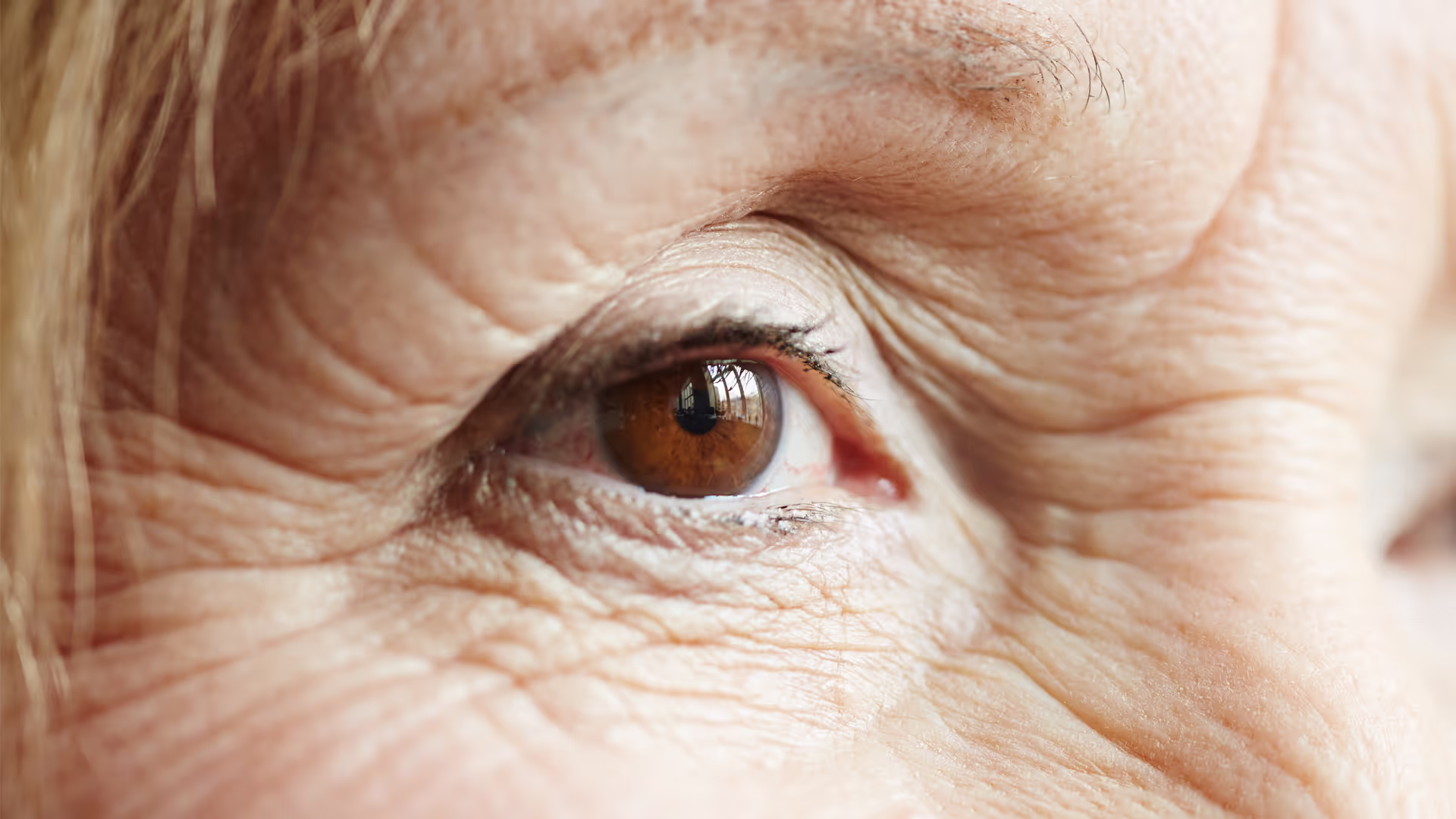
Third Eye Diagnostics is developing a first-of-its-kind device for the non-invasive measurement of ICP using the eye as a window to the brain. The process involves taking a video of the retina during gentle and brief contact of the instrument optical window with the cornea. This is used to determine the ocular pressure at which retinal vessel collapse occurs, this being a proxy measurement of ICP.
The optical challenge is to co-design multiple different imaging systems for integration into a handheld instrument format. Previous development prototypes had separately demonstrated the optics necessary for retinal and corneal video acquisition.
To obtain higher quality data, Third Eye Diagnostics now needed to add a new optical system to provide a fixation target for the patient to look at and stabilise the eye, alongside improved retinal and corneal imaging systems.
All of these systems needed to be designed, built and tested as a single sub-unit that could be retrofitted into Third Eye Diagnostics’ handheld prototype. To release product development funding, it was vital to demonstrate all the functionality in one device – something that had never been done before.

The highly talented and creative team at TTP rapidly and cost-effectively overcame several development hurdles leading to a medical device prototype for non-invasive management of hydrocephalus and traumatic brain injury. Their experience in the ophthalmic field enhanced their understanding of our objectives and resulted in creative solutions to complex optical and opto-mechanical design issues.
Terry Fuller
CEO, Third Eye Diagnostics
TTP’s optical designs deliver reliable patient data – and unlock potential new use cases for Third Eye Diagnostics’ technology
TTP substantially simplified the design of the existing retinal and corneal imaging systems, so that the additional optical path for a fixation target could be added using freed-up space in Third Eye Diagnostics’ prototype.
During the project, it also became apparent that the measurement system needed to account for natural variability in eye anatomy, and TTP’s experts proposed and developed an additional novel integrated optical system to overcome this challenge for Third Eye Diagnostics.
TTP’s optical design work included the following highlights:
- Simplification of Third Eye Diagnostics’ existing retinal imaging design from 6 complex aspheric lenses to a 3-lens design without loss in optical performance.
- Re-use and integration of Third Eye Diagnostics' existing retinal illumination system.
- Design of a reflection-based co-axial illumination and imaging system that collects corneal contact information.
- Design and improvement of a co-axial fixation target to enable the correct area of the retina to be measured, including simple switching between left and right eye targets.
- Optimisation of the use of off-the-shelf wavelength selective filters to enable all optical paths to be combined without significant optical losses that reduce retinal video SNR.
- Reduction of ghost reflections below noise levels from lens surfaces using non-sequential scattering simulation design so that reflections do not interfere with retinal video quality.
- Integration of the five optical paths into a single mechanical chassis that bolts directly into Third Eye Diagnostics' existing casework.
- Support for Third Eye Diagnostics during the integration and testing process.
In addition to the optical design work, TTP created new industrial and ergonomic concept designs for Third Eye Diagnostics’ handheld intra-cranial pressure monitor. The purpose of these was to improve the usability of the handheld device, but also to demonstrate how the new optical design can be integrated into an attractive instrument that opens up new use cases – including home use applications – for Third Eye Diagnostics’ technology.
About TTP's Ophthalmology and Optometry consulting team
Building on 30 years of pioneering advancements in ophthalmology, we have collaborated with the industry’s top companies to create sector-defining products. Our deep domain expertise, paired with our unrivalled agility in execution, ensures we consistently deliver high-impact results for our clients. From novel optics for contact lenses and IOLs, to innovative drug delivery and surgical systems, we are always looking for ways to address patient, healthcare professional and industry needs. Find out how our team of ophthalmology technology experts can help you.
TTP's Ophthalmology and Optometry team is part of a broader Medical Device Consulting business unit. Read more about TTP's approach to medical device design and development.